










Live. Laugh. Smile.
Teens
Impacted Teeth
There are many occasions in which a pre-teen or teen patient will come in for a consultation only to find out that they have impacted teeth. This means that one or some of their adult teeth are “stuck” in the bone because of minimal space for teeth to come in or due to issues with anatomical or biological eruption of teeth themselves.
Prevention of Impacted teeth
The teeth that show the highest prevelance of impaction are usually the upper canines (the third teeth from the centre). These teeth usually erupt into the mouth between age 10-11. At this stage if the primary or baby canines are not loose or have not fallen out, a routine panoramic x-ray should be taken. This x-ray gives us a clue as to why there is a delay in eruption of the upper canines. Sometimes patients just have teeth that take some time to come into the mouth. If this is the case, most orthodontists will just wait and allow nature to take its course.
Most permanent teeth emerge into the mouth by resorbing the roots of their primary successors. The permanent teeth function like “Pac Man” and “chew” the roots of the baby teeth until there is nothing to hold them in the jaw. Most permanent teeth become impacted when there is a problem with this process and as a result, primary or baby canines are prophylactically extracted to encourage the adult canines to drop into place.
There are several indicators to determine whether primary canines should be extracted. Clinical indications includes:
Based on x-ray analysis, we will recommend extraction of the primary canines for the following reasons:
There are also clinical signs that things may not be developing properly. If extractions are required they will be completed by your dentist. More often than not, prophylactic extraction of primary teeth usually helps the adult canine to erupt into place on its own without any orthodontic intervention. This is because the extraction of the primary canine leads to a path of least resistance for eruption.
Once the primary canines are removed, the permanent canines usually change their course from erupting on an angle to erupting downward toward the space created by the extracted primary canine. If the primary canines are not removed, there is a risk for impaction of the permanent canines
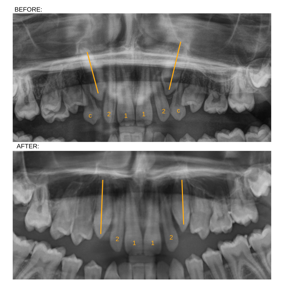
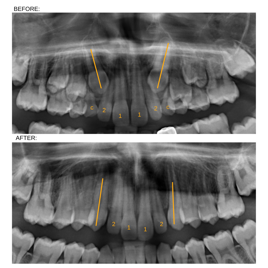
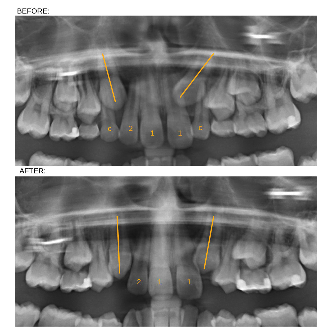
This x-ray shows the attributes of permanent canines that have a high chance of impaction. Eruption on an angle and sometimes past the roots of the lateral incisors (2), the roots of the primary canines (C) are not being resorbed or chewed away as the permanent canines due to their path of eruption. Sometimes a large bubble or eruption cyst is also seen on the crown of the canines (arrow).

Take a look at these cases showing the impact of prophylactically removing primary canines to prevent the impaction of the permanent canines:

Treatment of Impacted Teeth
Once teeth become impacted it is important to obtain a Cone Beam CT of the affected canine. This special test is a 3D-xray that allows clinicians to evaluate the position of the tooth in space. This helps in allowing the orthodontist to treatment plan how to bring the tooth into the mouth and will help the oral surgeon visualize and locate the tooth during surgery without removal of too much bone and tissue.
Once the exact position of the canine is corroborated, an expander may be placed first to help create space for the canine. Once the braces are bonded and the space is adequate to receive the impacted tooth in the correct position, the patient will be sent to the surgeon to uncover the tooth.
Uncovering or exposing the tooth is always completed by a surgeon or a periodontist. These professionals may use different means to help the patient relax during the procedure. These include, local anaesthetic alone or in combination with Nitrous Oxide Sedation (laughing gas) or conscious sedation (the patient is fully responsive to commands during this procedue but does not remember the procedure). The recovery from a surgical exposure lasts from a couple days to a week, and usually depends on the site of expsorue and how deep the canine was impacted.
During the surgery, the surgeon will uncover tissues, gums and surrounding bone from the tooth. If the tooth is impacted in the front of the bone closer to the lip, a gold chain will be bonded to assist the orthodontist in gradually pulling the tooth into the mouth. The tooth and gold chain are then covered with gum tissue that heals into place. This is referred to as a closed exposure. If the tooth is impacted in the palate of the mouth, and is not too deep, an open exposure is completed instead. This involved putting an islet or bracket to the tooth and covering the tooth with a periodontal dressing instead of suturing palatal tissue over. This way, the tooth is allowed to erupt into the mouth on its own.
The patient is usually seen by the orthodontist a week after the exposure is completed to start “tracking” the tooth in place. This may involve pulling the tooth into place with an elastic thread, wires or springs. It usually takes 2-3 years to complete a case in which teeth are impacted. The best way to prevent the occurrence of impacted teeth is to see your orthodontist by age 7 for an initial evaluation. Extraction of primary teeth and potential expansion of the upper jaw are ways in which the risk is further reduced.
Dr. Virdee has significant experience working with impacted teeth. If you may have an impacted tooth, feel free to call us for a consultation. Dr. Virdee will be happy to discuss your case and options with you.
Smile Gallery
-

Bringing an impacted canine into ideal alignment
Another case where our teenage patient had all their adult teeth present except the upper primary canines (c). An XRay showed that the upper right permanent canine was impacted in the palate. Once braces were bonded and enough space was created for the permanent canine (3), the primary canines were extracted and the permanent canines were exposed by a surgeon. The surgeon puts a dressing on the canines to prevent the tissue from overgrowing the impacted canines again. Braces are then used to bring the permanent canines into ideal position.
Total treatment time: 24 months
-

Bringing an impacted canine into ideal alignment
Another case where our teenage patient had all their adult teeth present except the upper primary canines (c). An XRay showed that the upper right permanent canine was impacted in the palate. Once braces were bonded and enough space was created for the permanent canine (3), the primary canines were extracted and the permanent canines were exposed by a surgeon. The surgeon puts a dressing on the canines to prevent the tissue from overgrowing the impacted canines again. Braces are then used to bring the permanent canines into ideal position.
Total treatment time: 24 months
-
Bringing impacted canines into ideal alignment
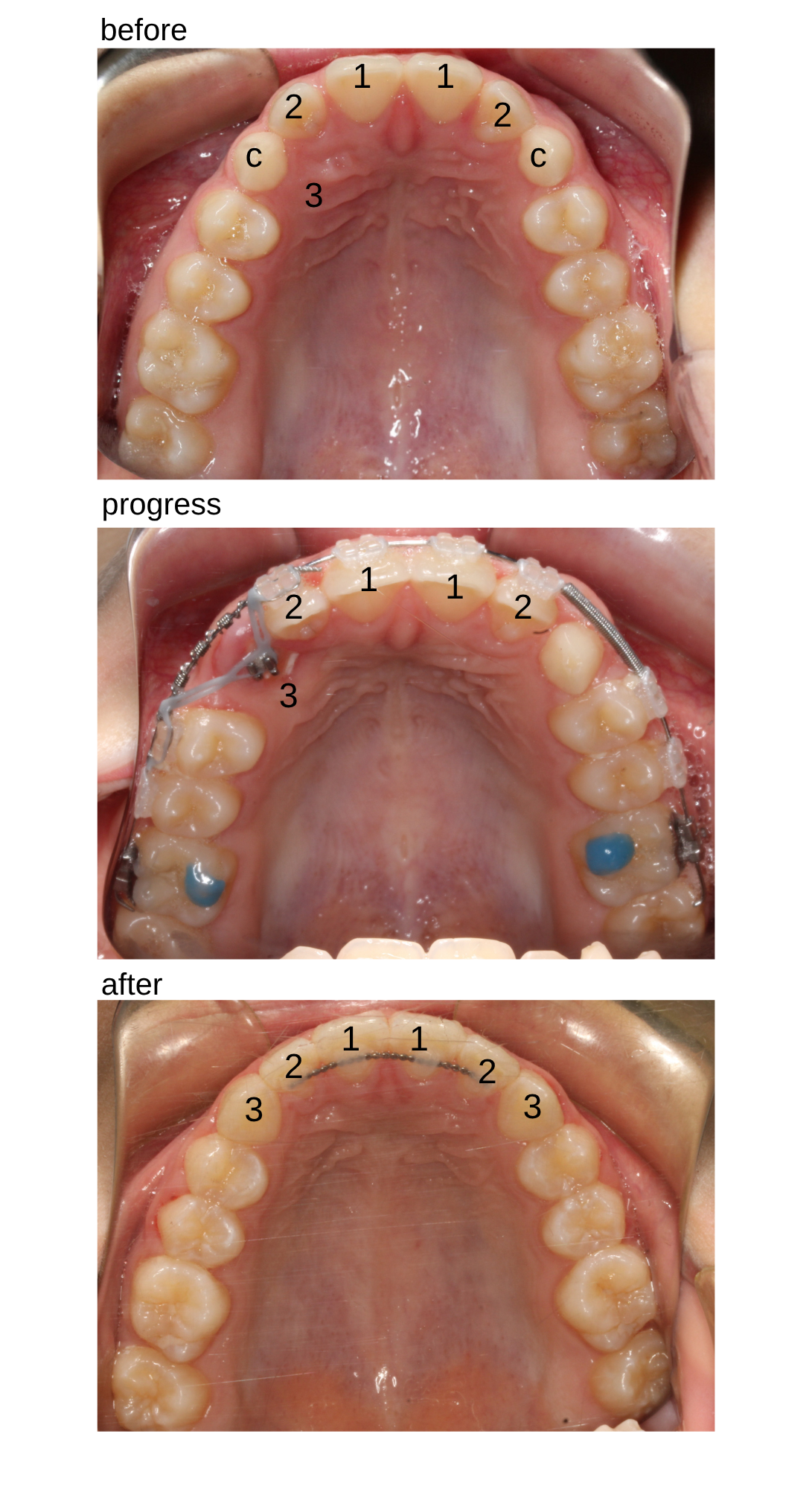
This 14-year-old patient had all their adult teeth present except the upper primary canines (c). An XRay showed that the upper right permanent canine was impacted in the palate. Once braces were bonded and enough space was created for the permanent canine (3), the primary canines were extracted and the permanent canines were exposed by a surgeon. The surgeon puts a dressing on the canines to prevent the tissue from overgrowing the impacted canines again. Braces are then used to bring the permanent canines into ideal position.
Total treatment time: 24 months
-

Bringing impacted canines into ideal alignment
This 13-year-old patient had all their adult teeth present except the upper primary canines (c). An XRay showed that the permanent canines were impacted in the palate. Once braces were bonded and enough space was created for the permanent canines (3), the primary canines were extracted and the permanent canines were exposed by a surgeon. The surgeon puts a dressing (right canine in photo) on the canines to prevent the tissue from overgrowing the impacted canines again. Braces are then used to bring the permanent canines into ideal position.
Total treatment time: 30 months


